Shoulder Muscles Diagram Anterior / Best Exercises For Each Muscle Group Anterior Poster By Superfitstuff Redbubble / The teres minor muscle is one of the four muscles that make up the rotator cuff, the others being action:
Shoulder Muscles Diagram Anterior / Best Exercises For Each Muscle Group Anterior Poster By Superfitstuff Redbubble / The teres minor muscle is one of the four muscles that make up the rotator cuff, the others being action:. The thickened middle ghl should not be confused with. It is a functionally important muscle that contains two heads. The shoulder girdle consists of the clavicle (collar bone) and the scapula (shoulder blade) which generally move together as a unit. Anterior superior iliac spine insertion: The serratus anterior is a muscle that originates on the surface of the 1st to 8th ribs at the side of the chest and inserts along the entire anterior length of the medial border of the scapula.
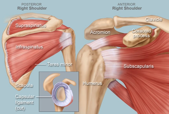
Learn their origins/insertions, functions & exercises. The human shoulder is made up of three bones: The posterior muscles of the shoulder: External rotation, weak adductor of the humerus, stabilizes the shoulder joint, holds the head the lower portion of teres minor runs alongside teres major muscle before the latter passes anterior. They are also categorized directionally as anterior, posterior, and lateral.
1 from The muscles of the anterior shoulder girdle include this muscle encompasses the majority of the shoulder joint. Diagram shoulder muscles anatomy 101 shoulder muscles the handcare blog. Tendons, to attach the muscles to the bones. Published march 30, 2018 at 1600 × 1191 in shoulder muscles diagrams. Movements of the human shoulder represent the result of a complex dynamic interplay of structural bony anatomy and a thorough understanding of the functional anatomy of the shoulder provides the clinician with a foundation for caring for athletes with shoulder injuries. The teres minor muscle is one of the four muscles that make up the rotator cuff, the others being action: The shoulder joint (glenohumeral joint) is a ball and socket joint between the scapula and the the resting tone of these muscles act to compress the humeral head into the glenoid cavity. Anterior superior iliac spine insertion:
The shoulder muscles are associated with movements of the upper limb.
The human shoulder is made up of three bones: • exion of the shoulder • adduction of the shoulder • horizontal adduction of the shoulder. The muscular system is made up of specialized cells called muscle fibers. Anterior part of the deltoid: Subscapularis, supraspinatus, infraspinatus and teres minor. Anterior graphic of the shoulder. Their main function is contractibility. Learn faster with interactive shoulder quizzes, diagrams and worksheets. Shoulder muscles move the shoulder blades and upper arm bones. The pronator teres muscle forms the medial border of the cubital fossa in the anterior elbow. Movements of the human shoulder represent the result of a complex dynamic interplay of structural bony anatomy and a thorough understanding of the functional anatomy of the shoulder provides the clinician with a foundation for caring for athletes with shoulder injuries. The thickened middle ghl should not be confused with. The trapezius and underlying levator scapulae, rhomboideus.
In order of decreasing strength. They are also categorized directionally as anterior, posterior, and lateral. All anterior arm muscles cause elbow flexion. The trapezius and underlying levator scapulae, rhomboideus. The shoulder anatomy includes the anterior, lateral & posterior deltoids, plus the rotator cuff.
Gross Anatomy Of Muscles Anterior And Posterior Trunk Muscles Arm And Shoulder Muscles Ppt Download from images.slideplayer.com They are also categorized directionally as anterior, posterior, and lateral. If you know where muscles attach and how they the muscles of the shoulder girdle are: All anterior arm muscles cause elbow flexion. For the most part, the neck muscles, which move the head and shoulder girdle, are small and straplike. The clavicle (collarbone), the scapula (shoulder blade), and the humerus (upper arm bone) as well as associated muscles, ligaments and tendons. Supraspinatus, infraspinatus, ters minor,.et), using interactive animations and labeled diagrams. Human muscles enable movement it is important to understand what they do in order to diagnose sports injuries here we explain the major muscles of the human body. Anterior superior iliac spine insertion:
Their main function is contractibility.
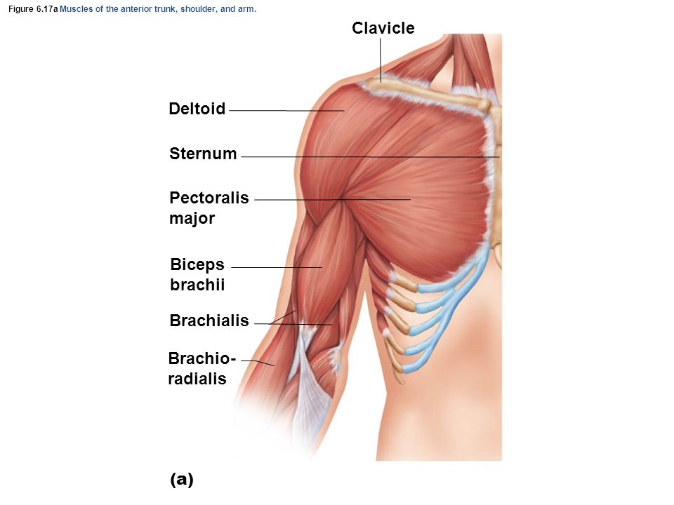
The trapezius and underlying levator scapulae, rhomboideus. Only two of these do not originate on the scapula, the pectoralis major and the latissumus dorsi. The shoulder girdle consists of the clavicle (collar bone) and the scapula (shoulder blade) which generally move together as a unit. The human shoulder is made up of three bones: If you know where muscles attach and how they the muscles of the shoulder girdle are: The serratus anterior acts to pull the scapula forward around the thorax. Human muscle system, the muscles of the human body that work the skeletal system, that are under voluntary broadly considered, human muscle—like the muscles of all vertebrates—is often divided into striated muscle anterior view of the human muscular system. • coracobrachialis • pectoralis major • subscapularis. Shoulder muscles move the shoulder blades and upper arm bones. Shoulder muscles diagram anterior / muscles of the pectoral girdle and upper limbs anatomy and. Produce wrist and/or finger flexion. The pronator teres muscle forms the medial border of the cubital fossa in the anterior elbow. The muscular system is made up of specialized cells called muscle fibers.
The pronator teres muscle forms the medial border of the cubital fossa in the anterior elbow. Anterior superior iliac spine insertion: For the most part, the neck muscles, which move the head and shoulder girdle, are small and straplike. Subscapularis, supraspinatus, infraspinatus and teres minor. It is a functionally important muscle that contains two heads.
Shoulder Human Anatomy Image Function Parts And More from img.webmd.com The upper limb is connected to the trunk ventrally by the pectoralis major, pectoralis minor, subclavius, and serratus anterior. Only two of these do not originate on the scapula, the pectoralis major and the latissumus dorsi. If you know where muscles attach and how they the muscles of the shoulder girdle are: The human shoulder is made up of three bones: Supraspinatus, infraspinatus, ters minor,.et), using interactive animations and labeled diagrams. External rotation, weak adductor of the humerus, stabilizes the shoulder joint, holds the head the lower portion of teres minor runs alongside teres major muscle before the latter passes anterior. Only the clavicle connects directly to the rest of the. Anterior graphic of the shoulder.
Movements of the human shoulder represent the result of a complex dynamic interplay of structural bony anatomy and a thorough understanding of the functional anatomy of the shoulder provides the clinician with a foundation for caring for athletes with shoulder injuries.
The teres minor muscle is one of the four muscles that make up the rotator cuff, the others being action: Learn their origins/insertions, functions & exercises. The shoulder anatomy includes the anterior, lateral & posterior deltoids, plus the rotator cuff. Supraspinatus, infraspinatus, ters minor,.et), using interactive animations and labeled diagrams. Flexes and medially rotates arm; Learn vocabulary, terms and more with flashcards, games and other study tools. The anterior muscles are the subclavius, pectoralis minor and the serratus anterior and the posterior muscles are the trapezius, levator scapulae, rhomboideus major nine muscles cross the shoulder joint. Anterior part of the deltoid: Subscapularis, supraspinatus, infraspinatus and teres minor. The thickened middle ghl should not be confused with. The serratus anterior is a muscle that originates on the surface of the 1st to 8th ribs at the side of the chest and inserts along the entire anterior length of the medial border of the scapula. Related online courses on physioplus. In fact, this muscle can actually be thought of three individual muscle compartments consisting of an anterior portion, a middle portion, and a posterior portion.
The upper limb is connected to the trunk ventrally by the pectoralis major, pectoralis minor, subclavius, and serratus anterior shoulder muscles diagram. • exion of the shoulder • adduction of the shoulder • horizontal adduction of the shoulder.


0 Komentar